
VMTK: Visual Manipulation Toolkit for NeuroImages
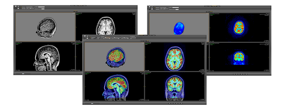
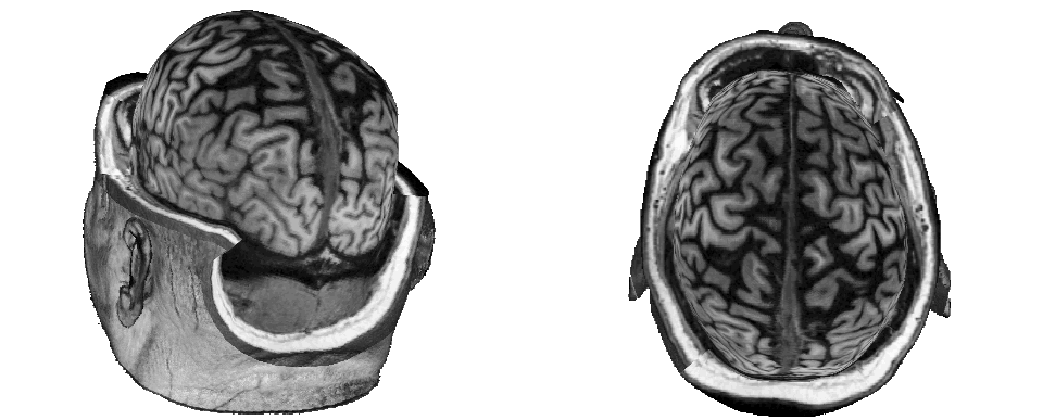
This project aims at implementing an interactive visualization tool which permits a physician to diagnose and to make pre-surgical planning for removal of cortical lesions. The cortical lesions are structural abnormalities (identifiable in original high-quality image data by a skilled neurologist) and seated in the outermost sheet of the brain. Features, as patient-oriented slice views, fused views of multimodalities, multiplanar reformatting and focus configuration, are presented in most medical applications -- in an independent, but not in an integrated, manner. This may hinder a thoroughly comparative visual analysis of multimodal exams. The key to our solution is:
1. to distinguish five reference spaces along the data processing from the raw data (in DICOM format) to the format of the displayed images. They are Patient-oriented coordinate reference (DCR), native coordinate reference (PhCR), normalized native coordinate reference (NPhCR), and texture coordinate reference (TCR).
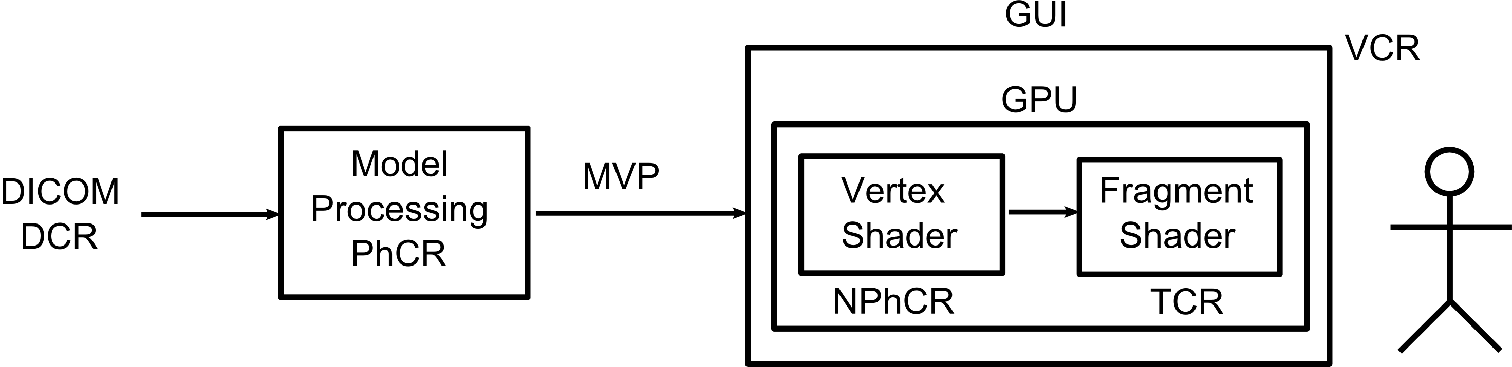
2. to move most of interactive visualization algorithms to GPU for reducing CPU to GPU data transfer latency. The raw data are loaded in the GPU memory with texture coordinates (TCR) and different views are derived directly from them on GPU. During user interactions only the updated control data Ω and the viewing data MVP are continually resent to GPU.